


Less commonly seen than superficial pyoderma, deep pyoderma breaks through hair follicles to involve the deep layers of the skin, resulting in furunculosis and cellulitis. Due to damage to blood vessels in the dermis, bloody discharge or haemorrhagic crusts are common with a risk of haematogenous spread and bacteraemia.
It may be preceded by superficial pyoderma that has not been managed effectively, but can also be manifested as local lesions, for example with callus pyoderma, chin pyoderma, interdigital nodules, and in a multifocal or generalised distribution. It can be seen with any underlying trigger or acquired immunodeficiency, but is commonly associated with demodicosis.
Clinical features
Lesions are variable and include serosanguinous to purulent fistulous tracts, haemorrhagic bullae, nodules and varying degrees of erythema and swelling, and may be painful. The presence of blood provides an important diagnostic clue to differentiate between deep and superficial pyodermas.
Pyodermas are almost invariably associated with underlying primary factors that need to be identified and treated for successful resolution. Lesions most often involve the trunk, pressure points (callus pyoderma, Figure 1), chin, nose and feet (interdigital pyoderma, Figures 2 and 3).
Post-grooming furunculosis, frequently involving Pseudomonas spp., is a newly recognised deep pyoderma that develops within a few days of shampooing and grooming.
In general, however, the most important causative bacterium, and also the most commonly isolated in deep pyoderma cases, is Staphylococcus pseudintermedius. Other species include S. schleiferi, S. aureus and occasionally gram-negative staphylococcal infections and anaerobic bacteria. Methicillin resistance is possible in all staphylococcal infections. Its increasing occurrence worldwide mandates culture and antibiotic sensitivity; if identified, rigorous hygiene measures and owner education are essential.


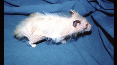
German Shepherd pyoderma is a poorly understood form of deep pyoderma, with likely genetic and immunological predispositions in German Shepherd dogs or their crosses (Figure 4). Treatment is often unrewarding, with relapse after cessation of treatment a common problem.
Predisposing factors include (after Hnilica and Patterson, 2017):
- Demodicosis, especially with pedal and facial pyoderma
- Allergic skin diseases
- Scabies
- Endocrinopathy (eg hypothyroidism, hyperadrenocorticism)
- Immunosuppressive therapy (particularly with glucocorticoids)
- Autoimmune and immune-mediated diseases, especially if there has been treatment with immunosuppressive drugs
- Trauma (eg callus pyodermas) and faulty conformation in some cases of pedal pyoderma
- Any chronic skin disease where the integrity of the skin defence mechanism is compromised
Differential diagnosis
- Demodicosis (an important common underlying cause; hair plucks, skin scrapes and in chronic cases biopsy are all advised)
- Allergic skin diseases
- Calcinosis cutis (lesions in chronic cases become secondarily infected, manifesting as deep pyoderma)
- Dermatophytosis (especially in chronic cases with secondary infection)
- Atypical bacterial infections such as actinomycosis or nocardiosis
- Autoimmune diseases
- Neoplasia

Diagnosis
Consider the history and clinical signs. There may be a history of superficial pyoderma that has not been effectively treated with subsequent deterioration and development of deep pyoderma. Rule out differential diseases. All cases will benefit from hair plucks, tape strips and skin scrapes.
Cytological examination is advisable in all cases. A suppurative to pyogranulomatous inflammation is a typical finding with phagocytosis of bacterial cocci and/or rods.
Culture and antimicrobial sensitivity is necessary in all cases. The primary pathogen is usually S. pseudintermedius but other bacteria may be found. Cytological examination with culture and antimicrobial sensitivity are best considered together to facilitate an accurate diagnosis. Disinfection of the outer part of a draining lesion followed by deep swabbing will reduce the risk of contaminant organisms. Where no discharging lesions are present, deep tissue may need to be obtained for culture by biopsy (the outermost part of the sample needs to be discarded before submitting in sterile saline).
Histopathological examination will reveal a deep suppurative/pyogranulomatous folliculitis, furunculosis, cellulitis and panniculitis. Intralesional bacteria may be difficult to find.
Clinical management
Deep pyoderma cases are challenging. Time spent with owners is essential before treatment and investigations are undertaken as the diseases are serious, require lengthy treatments and incur considerable expense.
Systemic therapy should only be administered based on culture and sensitivity and an evaluation of a cytological specimen. While guidelines specific for the treatment of deep pyoderma are still lacking, general recommendations of good antimicrobial stewardship should be followed (Beco et al., 2013). Antimicrobial therapy should be given at the full registered dose, and the dog should be weighed to ensure accurate dosing. A minimum of six to eight weeks of treatment is recommended, in general two weeks following clinical resolution. Close monitoring is critical and best done under veterinary supervision at all times, including cytological evaluation as clinical “cure” precedes absence of bacterial invasion.

Topical therapy can be instigated; in conjunction with systemic therapy, it will reduce surface contamination and support healing. Products should have proven antibacterial efficacy, such as those based on chlorhexidine. Underlying causes need to be investigated and treated. Topical therapy can be continued while these investigations are ongoing, and may be useful to prevent recurrence if no underlying cause is found.
When dealing with pyoderma cases, hand hygiene is very important and thorough disinfection of tables and instruments between cases is essential to avoid transmission of resistant and potentially zoonotic pathogens. This is especially important with multi-resistant pyodermas.
Multi-drug resistant pyodermas, like methicillin resistant S. pseudintermedius (MRSP), present a particular (and very difficult) challenge; in many cases, there will be no licensed antimicrobial agent possible. In this situation, specialist advice is recommended.


-1628179421-401x226.jpg)


